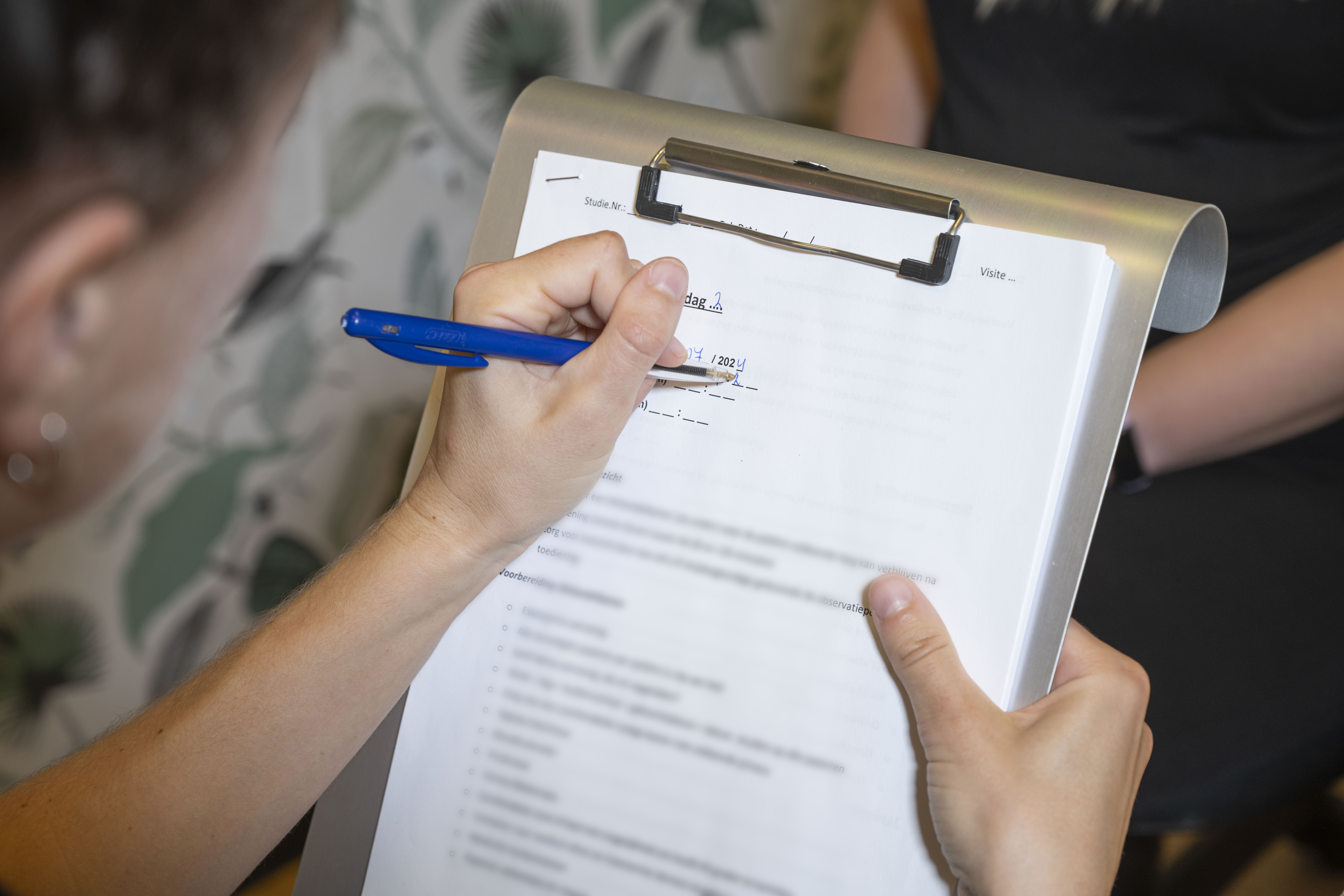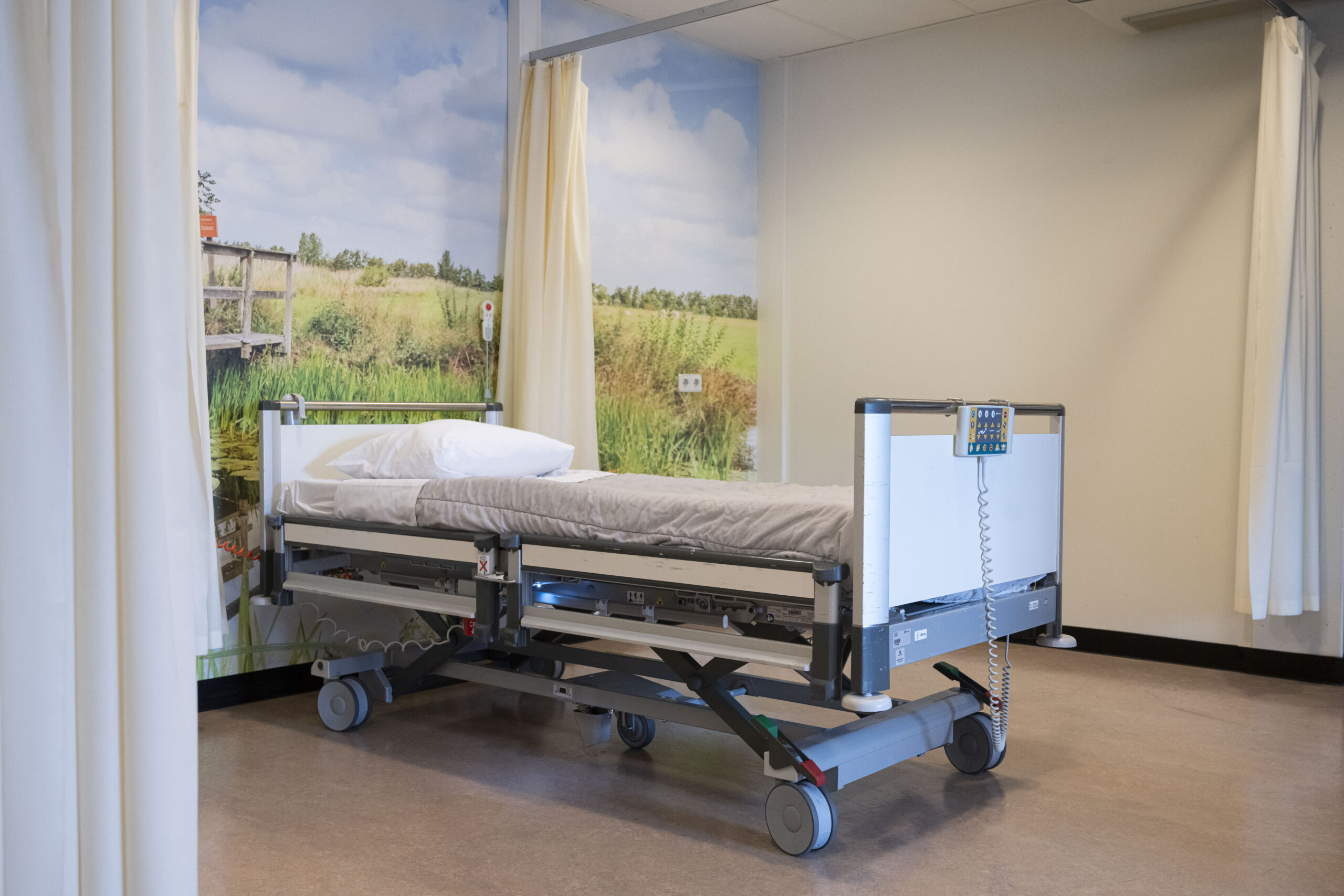
Photos of the medical equipment and the environment where ECT takes place.
Questionnaires are used to monitor the effects of the treatment.

This device is most commonly used to administer ECT. The dose can be read as a percentage (in the photo: 25%). On the paper on the right, brain activity (EEG) is also recorded.

These are the stickers placed on the head and body to measure brain activity.

These medications are administered through an IV. They induce anaesthesia and muscle relaxation.

These are two electrodes with gel that are held on the head. They deliver the electric current. There are also disposable versions that look like stickers..

This is a reusable mouthguard. Disposable versions also exist and look different. It is placed between the teeth once the patient is under anaesthesia to protect the teeth.

This mask delivers oxygen to the patient.

This is an example of a hospital bed in the recovery room where someone wakes up after ECT.

These photos are illustrative. The exact appearance of equipment and treatment areas may vary between hospitals and care facilities, but they give a general idea of what the ECT environment looks like.


